MtoZ Biolabs provides the Spatial Phosphoproteomics Service by integrating spatial partitioning strategies with phosphoproteomics analysis to systematically identify and quantitatively analyze protein phosphorylation states within specific spatial regions of tissues or cells. This service enables researchers to investigate region-specific regulatory features of signaling pathways while preserving spatial context, providing reliable data support for mechanistic studies and functional interpretation.
Principle of Spatial Phosphoproteomics
Spatial phosphoproteomics is a class of research strategies that builds upon spatial proteomics and focuses specifically on the spatial distribution and dynamic regulation of protein phosphorylation. As one of the most important post-translational modifications, phosphorylation exhibits strong spatial and temporal specificity in processes such as signal transduction, cell cycle regulation, and stress responses. While bulk phosphoproteomics analysis can reveal global phosphorylation changes, it often masks localized regulatory differences between distinct tissue regions or cellular subpopulations.
Wu, M. et al. Proteome Sci. 2024.
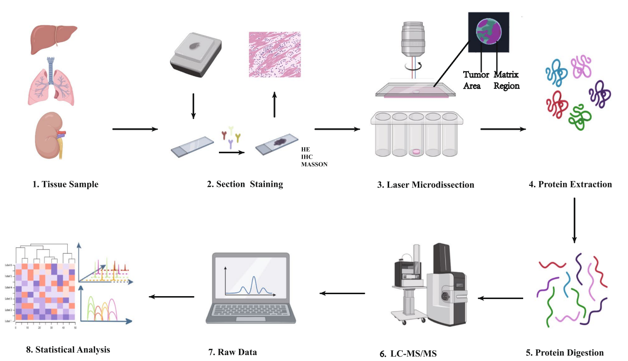
Figure 1. Spatial Proteomics
Spatial phosphoproteomics addresses this limitation by spatially partitioning tissues or cells and analyzing phosphorylated proteins and phosphorylation sites within defined regions while preserving positional information. This approach enables phosphorylation signals to be directly associated with tissue architecture, pathological regions, or local microenvironments and is particularly well suited for studying spatial differences in signaling pathway activation.
From a technical perspective, one major approach is region-based spatial phosphoproteomics analysis. This strategy typically begins with tissue sections, where regions of interest are defined through histological staining or microscopic observation, followed by precise sampling using laser capture microdissection (LCM) or microregion-level cutting strategies. Spatially confined samples are then subjected to protein extraction and enzymatic digestion, followed by phosphopeptide enrichment using strategies such as metal affinity–based or selective material–based methods. Subsequent LC-MS/MS analysis enables identification and quantification of phosphorylated proteins and sites, allowing spatially resolved characterization of phosphorylation regulation.
Another important approach involves the use of mass spectrometry imaging (Mass Spectrometry Imaging, MSI) to visualize the spatial distribution of phosphorylation-related signals. By acquiring mass spectrometric data through point-by-point scanning of tissue sections, MSI provides in situ information on spatial distribution trends of phosphorylated molecules or peptides, guiding downstream region-targeted analyses. Because phosphosite identification and quantification require high sensitivity and depth, MSI results are commonly combined with LC-MS/MS–based spatial dissection analyses to balance spatial visualization with robust phosphoproteomic characterization.
In practical applications, spatial phosphoproteomics often relies on multi-technology combinations to achieve an optimal balance among spatial localization accuracy, phosphosite coverage, and data interpretability. These integrated strategies enable systematic investigation of signaling pathway activation and regulatory differences across spatial regions.
Spatial Phosphoproteomics Service at MtoZ Biolabs
1. Region-Based Spatial Phosphoproteomics Analysis
MtoZ Biolabs defines specific regions within tissue sections through histological evaluation and microscopic localization and applies laser capture microdissection (LCM) or microregion-level cutting strategies to obtain spatially confined samples. Combined phosphopeptide enrichment and LC-MS/MS analysis are then used to systematically identify and relatively quantify phosphorylated proteins and phosphorylation sites across different spatial regions, enabling comparison of signaling pathway activation between tissue compartments or pathological areas.
2. Mass Spectrometry Imaging–driven Spatial Phosphorylation Distribution Analysis
Mass spectrometry imaging is applied to tissue sections using point-by-point scanning to construct spatial distribution maps of phosphorylation-related molecules. This approach supports the analysis of overall distribution patterns and region-specific phosphorylation signals and provides guidance for subsequent targeted spatial phosphoproteomics studies.
3. Integrated MSI and LC-MS/MS-Based Spatial Phosphoproteomics Strategies
Spatial distribution information obtained by MSI is combined with LC-MS/MS–based phosphoproteomics analysis of spatially dissected samples to enhance phosphosite identification depth and quantitative reliability while preserving spatial context, enabling complementary validation of spatial trends and molecular-level information.
4. Multiscale Spatial Phosphorylation Signal Analysis
Phosphorylation analysis can be performed at the tissue level, microregion level, or finer spatial scales, allowing systematic characterization of signaling pathway regulation across multiple spatial hierarchies.
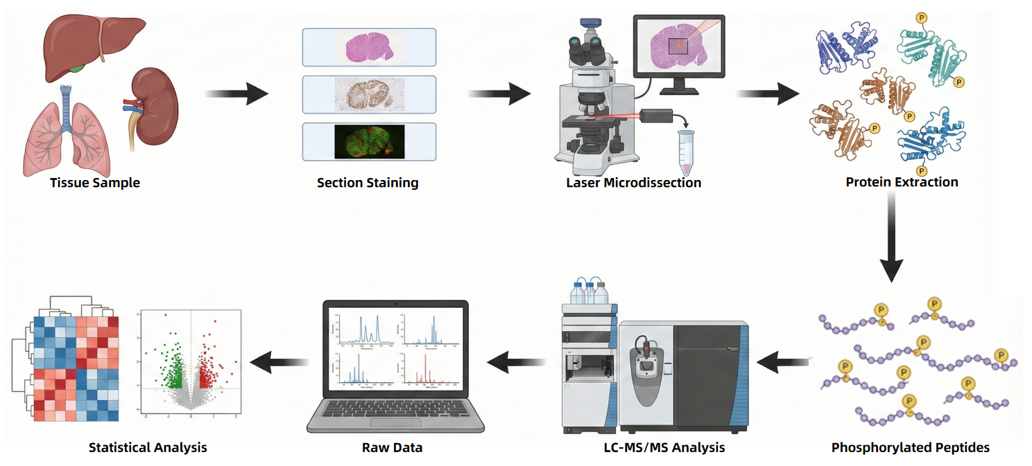
Workflow of Spatial Phosphoproteomics Service
Why Choose MtoZ Biolabs
- Experience in projects integrating phosphoproteomics with spatial analysis
- Support for multiple tissue types and diverse spatial partitioning strategies
- Mature and stable mass spectrometry workflows with high reproducibility
- Clear communication, standardized service processes, and transparent pricing
Applications of Spatial Phosphoproteomics Service
- Analysis of signaling pathway activation across different tissue regions
- Spatial regulation of phosphorylation in tumor microenvironments or pathological areas
- Investigation of region-specific phosphorylation changes during development
- Signal regulation studies in the nervous system and structurally complex tissues
- Exploration of spatial regulatory mechanisms of key kinases and their substrates
Start Your Project with MtoZ Biolabs
Contact MtoZ Biolabs to discuss your research objectives and sample types with our technical team and advance your Spatial Phosphoproteomics research project.
FAQs
Q1: What types of samples are suitable?
This service is suitable for a wide range of tissue or cell samples, particularly those with defined spatial structures or regional signaling pathway differences. Specific sample types can be confirmed prior to project initiation.
Q2: What is the service's general workflow?
Q3: What data formats are provided?
MtoZ Biolabs provides multiple standardized data formats to ensure that results can be efficiently used for downstream data processing, interpretation, and visualization. Standard deliverables include:
-
Raw mass spectrometry data files generated from LC-MS/MS platforms for phosphoproteomics analysis
-
Processed data tables (CSV or Excel format) containing identified phosphorylated proteins, phosphorylation sites, and relative quantitative information, annotated by spatial region or sampling location
-
Summary reports in PDF format, including analytical workflows, phosphopeptide enrichment information, quality control results, and key findings
-
Figures and visualization files related to spatial phosphoproteomics analysis, such as region-specific phosphorylation profiles or comparative plots, provided in high-resolution image formats (TIFF or PNG)
Customized data outputs can be provided upon request to meet specific data structure or publication requirements.
Q4: How should I prepare my samples?
To ensure accurate and reproducible results for the Spatial Phosphoproteomics Service, MtoZ Biolabs recommends preparing samples according to the following guidelines:
- Sample Type: The service supports a wide range of tissue- or cell-derived samples, including fresh-frozen tissues, tissue sections suitable for spatial analysis, cell pellets, and other samples with clearly defined spatial or regional characteristics.
- Sample Purity: Samples should be free from excessive contaminants such as particulates, high concentrations of salts, detergents, or strongly interfering chemical additives that may compromise phosphopeptide enrichment efficiency or mass spectrometry detection.
- Sample Amount: Sufficient sample material should be provided to support spatial partitioning or microdissection and subsequent phosphopeptide enrichment and mass spectrometry analysis. Specific input requirements can be confirmed in advance based on tissue type, spatial resolution, and study design.
- Storage and Shipping: Biological samples should be stored at −80°C to preserve phosphorylation states and shipped on dry ice to ensure sample integrity during transport.
- Sample Documentation: Please provide comprehensive background information, including sample source, tissue type, regions of interest, preparation methods, buffer composition, and the intended scope of phosphoproteomics analysis.
For more information, please refer to Sample Submission Guidelines for Proteomics and Sample Submission Guidelines for Metabolomics.